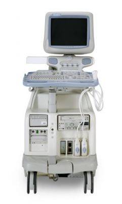
GE Healthcare Ultrasound Machine Rental Boston Massachusetts
This company is a nationwide service provider.

More by Standard Ultrasound
Ultrasound Description
Business to Business Only
Massachusetts Diagnostic Equipment Ultrasound Machine Rentals Boston Medical Devices For Rent
Cardiac Ultrasound Machine rentals in Boston, MA. The Vivid 7 Dimension ultrasound machine delivers more 4D imaging capabilities than ever before - ungated, unspliced, online and in real time or 4D full volume acquisitions. Vivid 7 is more than an ultrasound product. It's a way of thinking. Of working. A raw-data ultrasound platform that evolves. Renews. Improves. Year after year.
The GE Vivid 7 Ultrasound features can include: Colorflow Digital Ultrasound System w/THI, w/DICOM, w/3D View with B, B/M, M Mode. TruScan, TruSpeed Color Flow, Real Time Anatomical M-mode, Tissue Synchronization Imaging, Strain Rate Imaging, Real Time Full Volume, Tri-Plane Imaging, Tissue Velocity Imaging, Coded Phase Inversion for Contrast Imaging, Blood Flow Imaging (BFI)
Applications
- Cardiology
- Vascular
- Abdominal
- Small Parts
- MSK
- Urology
- Pediatric
- Neonatal
Comparable Systems
- Philips iE33
- Toshiba Aplio
- HP5500
Key Standard Features
- B, CFM, M, PW, PDI
- Anatomical M-Mode
- 17’’ High-Resolution TFT LCD Screen
- Tissue Harmonics
- Coded Harmonic Imaging
- Coded Excitation
- Anatomical M-Mode
- Auto Tissue Optimization (ATO)
- Auto CFM Optimization (ACO)
- Auto Spectrum Optimization (ASO)
- TruScan Architecture (TruAccess, SmartScan, ComfortScan)
- Internal DVD/CD-RW Drive
These Cardiac Ultrasound Machines are used to perform Echocardiogram's, often referred to as cardiac ECHO or simply an ECHO, it is a sonogram of the heart. Also known as a cardiac ultrasound, it uses standard ultrasound techniques to image two-dimensional slices of the heart. The latest ultrasound systems now employ 3D real-time imaging.
In addition to creating two-dimensional pictures of the cardiovascular system, an echocardiogram can also produce accurate assessment of the velocity of blood and cardiac tissue at any arbitrary point using pulsed or continuous wave Doppler ultrasound. This allows assessment of cardiac valve areas and function, any abnormal communications between the left and right side of the heart, any leaking of blood through the valves, and calculation of the cardiac output as well as the ejection fraction. Other parameters measured include cardiac dimensions and E/A ratio.
Echocardiography is performed by cardiac sonographers, cardiac physiologists or doctors trained in cardiology.
Click to Call Standard Ultrasound
Related Rental Listings


